Swiss Mummy Dental Project
The SMDP is a sub-project of the
SMP and deals with the description and analysis of dento-alveolar
pathologies of mummies mostly from Swiss collections. In some cases, the front
teeth are directly visible and can provide information about the health
of the individual. For the comprehensive examination of the oral cavity
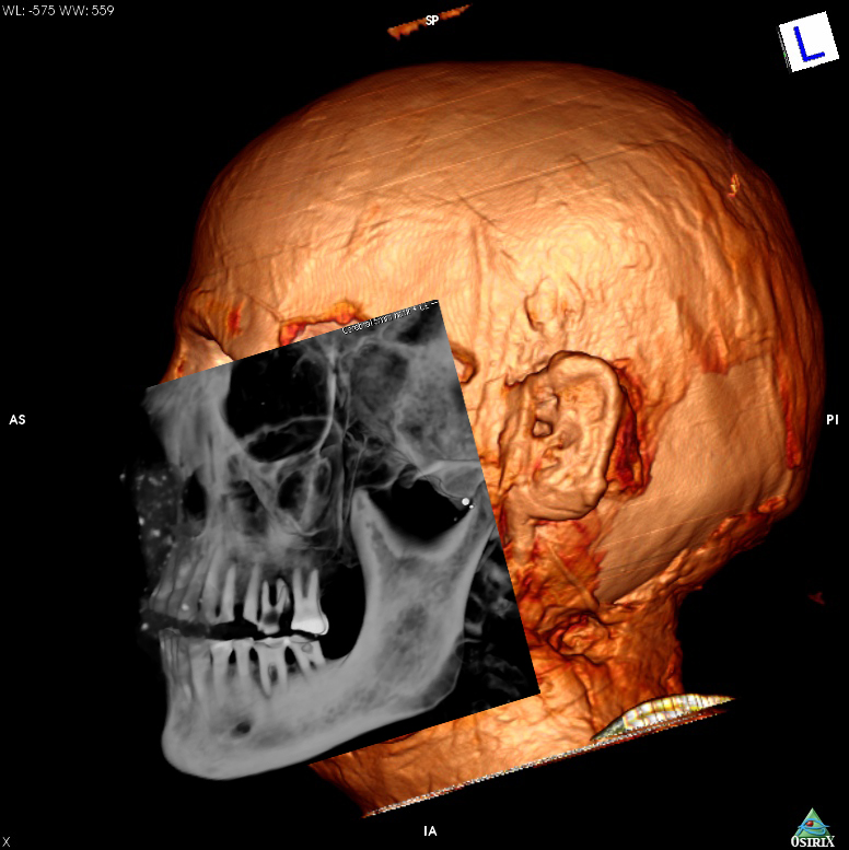
and skull, it is usually relied on radiological technology of CT-scans. In CT
scans with good resolution abrasion of teeth, which is very
frequently seen in ancient Egyptian mummies, but also carious lesions, consequences
of periodontitis or infection of the jaw are visible.
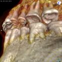
Schepeneses and Neshous teeth
If Schepenese would have lived longer, the progressing abrasion in addition to developing caries would probably have led to serious problems, as in the case of the about 50 year old Neshou (Musée d’ Yverdon et Région, Yverdon, Inv.Nr.: MY/3775). In this mummy, s deep carious lesion and the massive dental abrasion caused a severre infection around the roots of an upper molar.
Read more about caries here!
Read more about Neshou here!
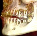
View the reconstruction of the dentition of Schepenese !
The film, based on a three-dimensional reconstruction of tomographic sections, first rotates the mummy skull. The cranial sutures are visible from above. In the sectional view the eye sockets appear first, then the base of the skull, the nasal cavity and finally the upper and lower jaw. Then follows a tour through the oral cavity. Except for the wisdom teeth, all teeth are present and caries-free, but quite worn – a finding that was widespread in ancient Egypt due to abrasive foods.